“Perhaps the most remarkable features of the molecule are its complexity and its lack of symmetry. The arrangement seems to be almost totally lacking in the kind of regularities which one instinctively anticipates, and it is more complicated than has been predicted by any theory of protein structure.”
— John Kendrew, describing the first structure of myoglobin in 1958
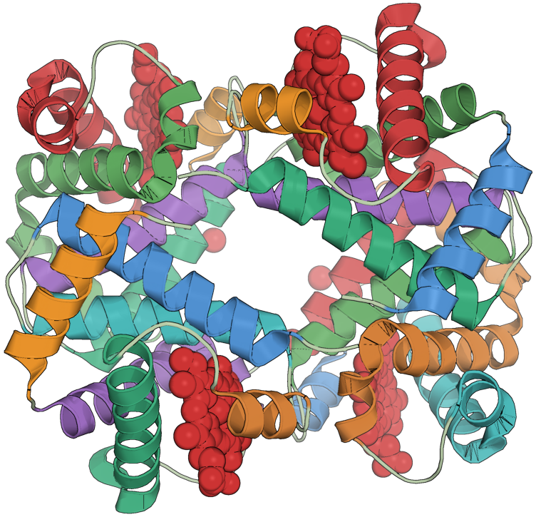
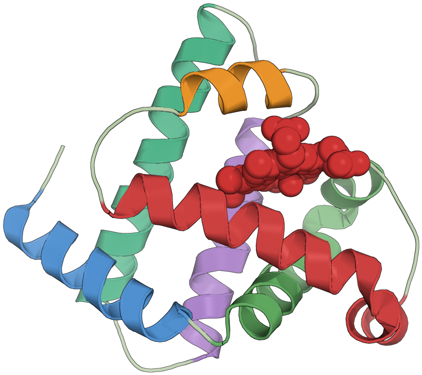
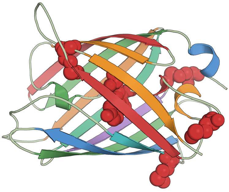
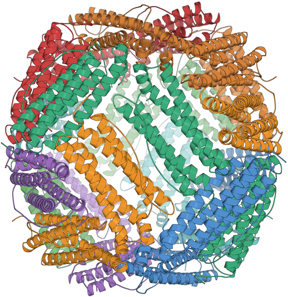
For the longest time, I’ve found proteins to be deeply compelling and yet at the same time extremely elusive. After some time in drug discovery, I’ve come to understand my feeling for them. Yes, proteins are fundamental as they are the molecular workhorses of life. And yes, they are essential for understanding disease and subsequently developing therapeutics through drug discovery and drug development. The study of proteins also allow us to understand biology itself. The list goes on and each in their own form is compelling enough for why one might want to study proteins and perhaps even dedicate a lifetime of work to them. Here, I will take a different route. Proteins exhibit a unique aesthetic quality through their structure. It’s quite amazing that proteins on one hand exhibit such tightly constrained structures shaped by evolution, but on the other hand are so irregular, flexible, and dynamic that they have the capability to handle all the necessary functions required for life. Here, it is also necessary to highlight that our ability to appreciate this aesthetic is only made possible by the efforts of the protein scientists, who have dedicated their careers to solving their structures (Figure 1). In what follows, I walk through the basic levels of protein structure in the hope of better understanding and appreciating both how proteins are organized and why their forms are so remarkable.
Structural Hierarchy of Proteins
This discussion draws primarily from Chapters 1–5 of Introduction to Protein Structure by Branden and Tooze. All ball-and-stick diagrams are reproduced from this source. I highly recommend the book to anyone seeking a deeper understanding and appreciation of protein structure.
Primary Protein Structure

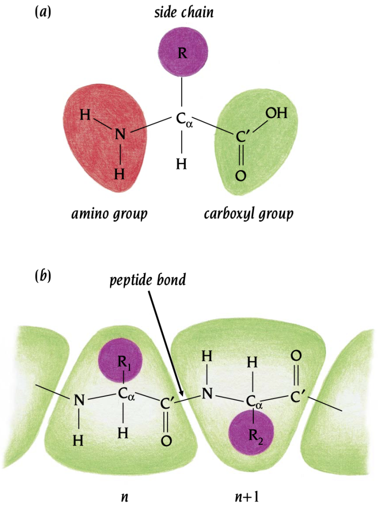
At its primary structure, proteins are quite simple. They are a sequence of amino acids, where each amino acid is one of 20 of the possible ones (Figure 2). Figure 3a depicts the structure of a single amino acid, where a central carbon atom is connected to an amino group, carboxyl group, side chain, and hydrogen. Figure 3b displays peptide bonds connecting amino acids to form a polypeptide chain. The repeating amino acids are called residues and are divided into the main chain atoms, identical in all residues, and the side chain atoms, different for different residues. While this representation is complete, a protein is not only a sequence, and its amino acids orient in 3D space, leading to a much more complex secondary structure.
Secondary Protein Structure
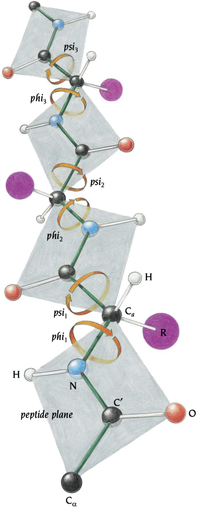
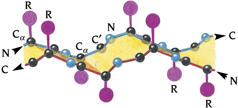
Secondary structure is tightly constrained, and all of the degrees of freedom for each amino acid can be summarized by a collection of torsion angles, twists around bonds, involving four atoms. Figure 4 illustrates a polypeptide chain and torsion angles at each residue responsible for folding, denoted phi and psi. Here, main chain atoms are grouped into peptide groups shown in gray. These peptide groups are approximately planar and rigid, leaving bond lengths and angles unchanged across all residues. Hence, a protein’s structural degrees of freedom lie nearly completely in its phi and psi torsion angles.
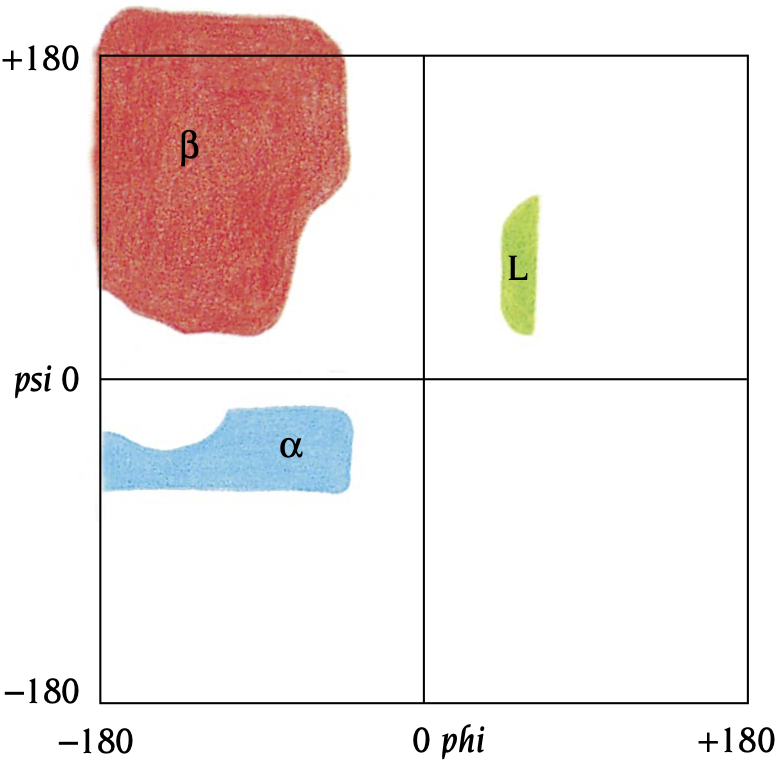
Interestingly, most combinations of phi and psi are forbidden due to collisions between an amino acid’s side chain and its main chain. In fact, the possible combinations can be theoretically computed according to these geometric constraints. Biophysicist G.N. Ramachandran is responsible for this idea and a Ramachandran plot (Figure 5) illustrates the distribution of allowed torsion angle pairs.
Alpha Helices and Beta Sheets
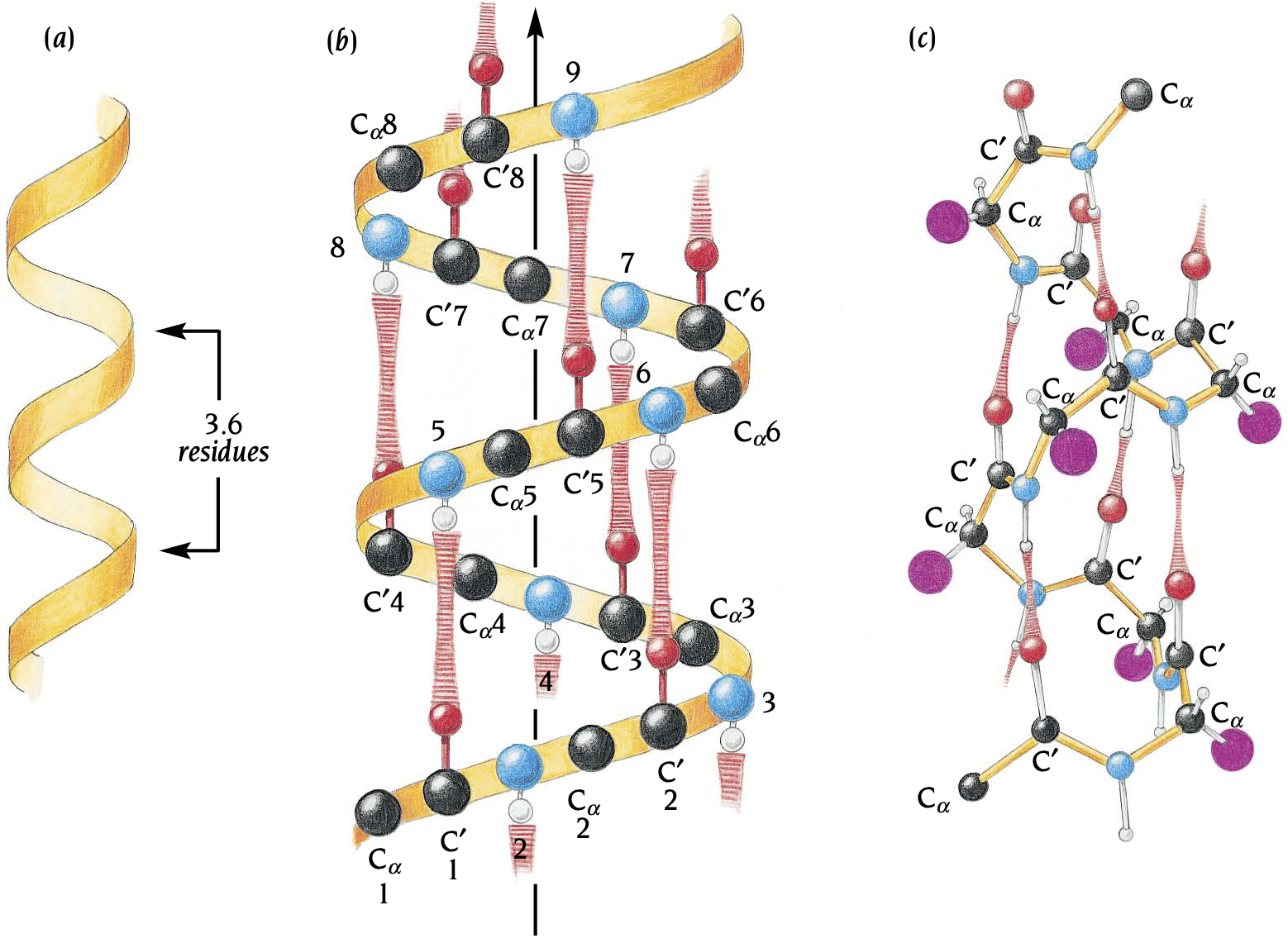
Folding is largely driven by hydrophobic (water-fearing) and hydrophilic (water-loving) interactions. A protein folds to pack its hydrophobic side chains into the interior of the molecule, forming a hydrophobic core, while orienting its hydrophilic side chains on its exterior creating a hydrophilic surface. This phenomenon however presents a challenge for proteins: in order to pack hydrophobic side chains into the interior, the hydrophilic main chain must also fold into the interior. The solution to the problem is quite an elegant one in secondary structures involving alpha helices and beta sheets (Figure 6). Alpha helices are a stretch of consecutive residues each involving phi and psi angle pairs of approximately 60 to 50, adopting angles in the bottom left quadrant of Figure 5. Importantly, the alpha helix exhibits 3.6 residues per turn, enabling hydrogen bonds between the th residue and the 4 residue. In other words, the helix structure satisfies the hydrophilic nature of the main chain, while the hydrophilic side chains can orient on the exterior and the hydrophobic side chains can pack on the interior.
Beta sheets exhibit a similar principle, but are structured as strands (Figure 7). They adopt phi and psi angles in the upper left quadrant of Figure 5. As they are oriented in strands, hydrogen bonding occurs between the main chains of pairs of strands, while the side chains orient above and below the main chains. Most of protein structures are formed by combinations of alpha helices and beta sheets, connected by loops.
Motifs
Combinations of secondary structures form motifs. These are frequently recurring patterns of secondary structure with precise geometric orientation. Each of these can be associated with a clear function such as DNA binding or have no specific function alone and are instead a part of the larger structural organization. The simplest ones of them are the helix-loop-helix (Figure 8a), two alpha helices joined by a loop, and the hairpin beta motif (Figure 8b), two antiparallel strands joined by a loop.
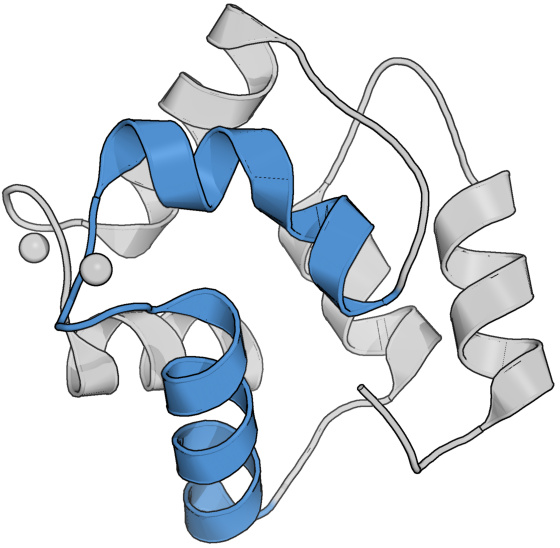
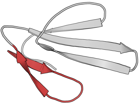
Figure 8. (a) EF hand motif in muscle protein, parvalbumin. (b) Hairpin motif in structure of snake venom, erabutoxin.
Tertiary Protein Structure
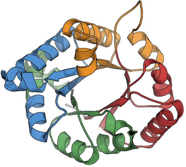
Combinations of motifs form domains, the primary unit of tertiary structure. Domains are defined as parts of the polypeptide chain (or the entire chain) that can fold independently into a stable structure, and unlike motifs, domains are also always associated with function. Fig. 9 illustrates how four consecutive beta-alpha-beta-alpha motifs form into a single domain comprising the structure for the metabolic enzyme, Triose Phosphate Isomerase.
And that concludes this brief tour through protein structure! Below, I’ll leave the reader with a few additional examples that, to me, capture some of the beauty of proteins.